


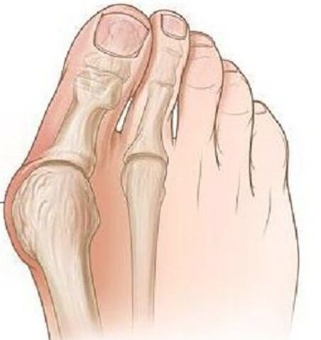
The valgus deformation of the thumb of the foot is the most common deformation of the foot, which is characterized by the deviation of the thumb.
Pathology is bilateral nature and mainly affects women over 35 years.In most cases, the deformation is accompanied by chronic inflammation of the joint bags and the deformation of the arthrosis of the plusnephalang joints, pronation deviations of the first midfoot bone.
Causes of the disease.Why is it dangerous?
The etiological causes of the valve deviations of the foot base have not yet been completely examined.There are several theories of occurrence:
- Remaining theory.In the middle of the 19th century it was assumed that only models of this deformation were exposed to high halves due to the use of model shoes, but during the study this pathology was found in men who wore flat shoes.
- The theory of primary muscle weakness - was refuted in a detailed examination of the problem.
- The theory of the weakness of the band apparatus and the lack of aponeurosis of the sole - most scientists adhere to this theory.
There are a number of factors that make the debut of this pathology:
- Obesity increases the load on the feet.
- Age -related dystrophic changes in the ligamentous joint apparatus.
- Weekens of Heeled shoes are more than 5 cm and are narrowed to the toe.
- The existing deformations of the skeleton (scoliosis, valgus deformation of the thigh and knee joints, flat feet).
The risk of pathology is that this deformation leads to a deformation of the attitude over time, and it is possible to develop dystrophic depth of the spine, the hip and knee joints, since improper distribution of the shop is accompanied by constant pain, which deviates the employment of humans.
Clinical manifestations
At the beginning of the disease, the valgus deformation is not clinically.Most women are more concerned about the appearance of the increase in the foot of the Metacarpal-Phalanx joint, and the outbreak of the first finger becomes noticeable.
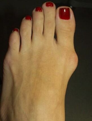
In the course of time, pain occurs, first when wearing narrow shoes and a persistent walking, and later the pain begins to be constant and hurts.
Due to the improper distribution of the weight, the limb is formed by the first and second fingers on the sole, the second finger is increased, strongly extended, the "Malleus" fingers, which are very complicated to walk.Due to the deterioration in blood circulation and innovation in the front of the foot, arthrosis and chronic bursitis develop.
Diagnosis
- During an objective examination, the classic deformation with valgus deviations in the thumb is visible, the distal part of the foot is expanded.When projection of the head of the first midfoot leg bone, there are signs of bursit - hyperemia and edema of the skin, pain during the palpations.
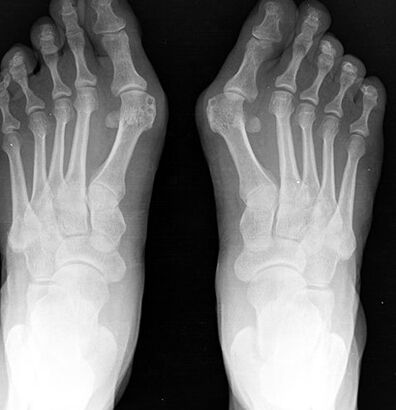
- The radiography of the foot in two projections.The front projection determines the degree of thumb, the condition of the Metacarpal-Phalanx joint, the degree of displacement of the sesamoid bone, and in the lateral projection, the degree of flat feet is made visible and calculated, which often occurs with valgus devastations of the first finger of the foot of the foot.
- Plantography.On the foot made on paper, a line is pulled through the middle of the heel, and between the fourth and third fingers the outer foot of the foot is formed in a figurative sense.In these patients, a flattened foot or flat feet of the 1st degree is more often found.
Treatment and prevention
Depending on the severity of the process, conservative or surgical treatment is carried out.
Conservative treatment is carried out in the mild stage of the disease.Different types of orthopedic insoles are used, which are selected individually and bring the deformed finger into the normal position, while the load is stabilized and distributed on the front of the limb.
In child and senile age, the crossbound of the distal extremity is used with a clearing between the first and the second finger.To reduce pain, warm baths with sea salt and soda are used, radon baths provide a good result.
Local anti -inflammatory gels, compresses with dimexid and lidocaine are used to combat bursitis, and Novocaine blockade and intra -articular injections of glucocorticoids are used with pronounced pain.
Surgical treatment is possible at every stage of the disease, it is carried out under local anesthesia, which reduces the list of contraindications for the operation.
In the 1st stage, the bone cold growth on the inner edge of the bone is removed, while it is only possible to reduce the pain of the process, but not to prevent the development of deformation in the future.
With a strong deviation of the finger with flat feet, an osteotomy on the base of the plus bone of the first finger is carried out using a bone blade to reject your finger into the normal position, with the help of a lavsan adhesive tape, the cross band of the sole is formed.After the operation, the leg is closely attached to a bandage for 4 weeks.It is recommended to use individual orthopedic insole in the course of the year.
The prevention of the disease is to use comfortable shoes without high paragraphs, shoes should sit freely on the leg, a narrowed toe should not bring discomfort in the thumb.If the skeleton is deformed, it is recommended to undergo preventive studies on the orthopedic surgeon in order to identify and slow down the progression of the valgus deformation of the foot.
Consequences and complications
A complication of the process is the development of dyke countries or the "march" of the foot, which is characterized by acute pain due to the micro -rises in the mating joints of the foot and tendovaginitis.
Due to the constant inflammatory process and the mechanical damage, there is a risk of developing malignant neoplasms of the bone.